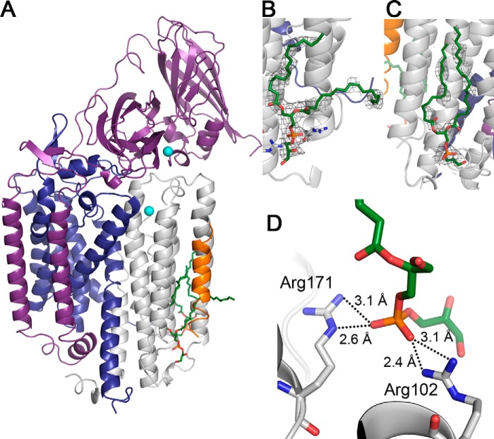
FIGURE 6.
Crystal structure of Methylocystis sp. str. Rockwell pMMO. A, one protomer shown with the pmoB, pmoA, pmoC, and mystery helix polypeptides in purple, blue, gray, and orange, respectively. Copper is depicted as cyan spheres, and lipids are shown as green sticks. B, zoomed-in view of the lipid-binding site located between the pmoC subunit and the mystery helix. The mystery helix and a pmoC helix consisting of amino acids 66–92 are omitted for clarity. C, zoomed-in view of the lipid-binding site located at the pmoC/pmoA interprotomer interface. The 2mFo − DFc composite omit map calculated with simulated annealing is superimposed over the lipids in both B and C in gray (1σ). D, hydrogen bonding interactions between the pmoC subunit and the lipid located at the site between pmoC and the mystery helix. The mystery helix is not shown in this panel. Nitrogen, phosphorus, and oxygen are colored blue, orange, and red, respectively.

