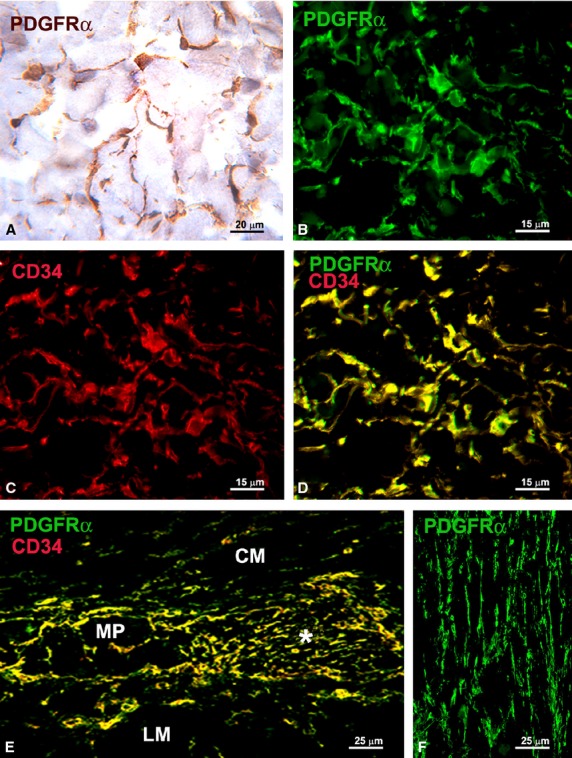
Fig. 1.

(A, B and F) PDGFRα-immunoreactivity; (C) CD34-immunoreactivity; (D and E) PDGFRα/CD34 double labelling. (A) Immunohistochemistry, haematoxylin counterstain; (B–F) Immunofluorescence. (A–D) Submucosa (stomach). PDGFRα-positive cells (A and B) and CD34-positive cells (C) form a 3-D network. All the PDGFRα-positive cells are also CD34-positive (D). (E) Myenteric plexus region (large intestine). PDGFRα/CD34-positive cells surround a ganglion (left side, MP) and form networks in the intergangliar region (right side, asterisk). (F) Circular muscle layer (small intestine). PDGFRα-positive cells form networks among the smooth muscle cells. CM: circular muscle layer; LM: longitudinal muscle layer. Scale bars are indicated in each panel.
