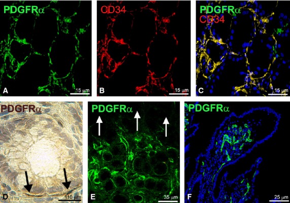
Fig. 3.

(A and D–F) PDGFRα-immunoreactivity; (B) CD34-immunoreactivity; (C) PDGFRα/CD34 double labelling. (A–C, E and F) Immunofluorescence (nuclei are blue stained with DAPI in (C and F); (D) Immunohistochemistry, haematoxylin counterstain. (A–C) Mucosa (stomach). Numerous PDGFRα/CD34-positive cells surround the funds of the glands. (D) Mucosa (large intestine). Few and scattered PDGFRα-positive cells surround glandular crypts (black arrows). (E) Mucosa (stomach). PDGFRα-positive cells are numerous around funds of the glands and absent from the superficial mucosa (indicated by white arrows). (F) Mucosa (small intestine). Only endothelial cells of capillary vessels display PDGFRα-immunoreactivity. Scale bars are indicated in each panel.
