Fig. 4.

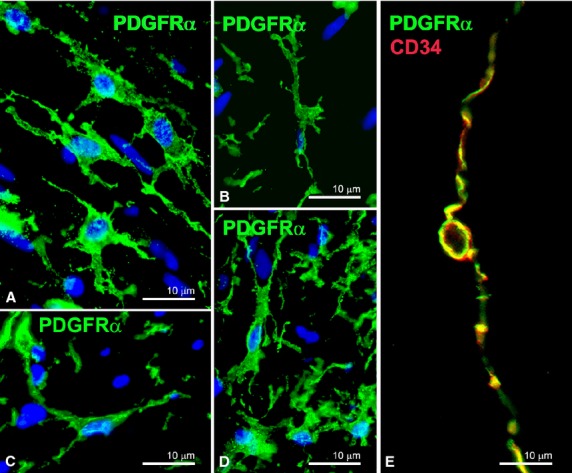
(A–D) PDGFRα-immunoreactivity (nuclei are blue stained with DAPI); (E) PDGFRα/CD34 double labelling. (A and B) Muscle layers (small intestine). Intramuscular PDGFRα-positive cells display two long telopodes and several short processes starting from the nucleated portion. (C) Submucosa (small intestine). PDGFRα-positive cells show a triangular body and three long and varicose telopodes. (D) Myenteric plexus region (small intestine). PDGFRα-positive cells display an oval body and several telopodes running in every direction. (E) A PDGFRα/CD34-positive cell at the border of a circular muscle bundle (small intestine) shows a small nucleated body and two long and thin telopodes starting from the opposite poles of the cell and with podomers and podoms clearly identifiable. Scale bars are indicated in each panel.
