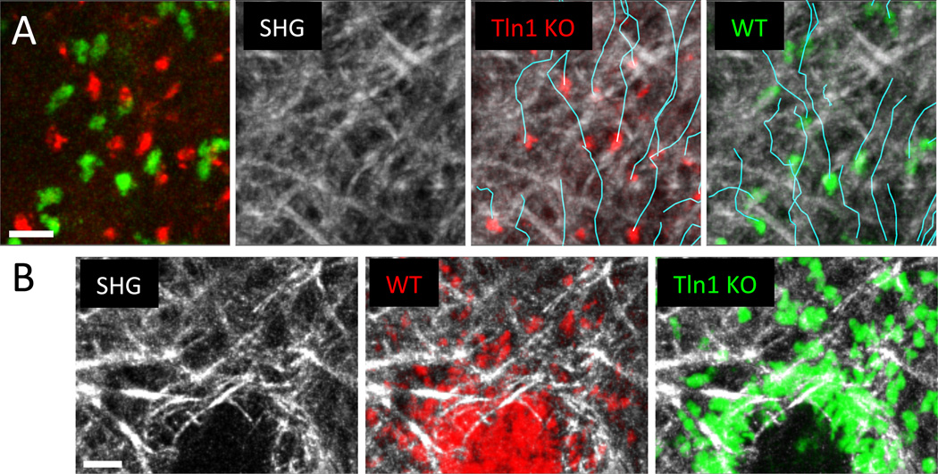
Fig. 3. Nonadhesive neutrophil migration in the dermal interstitium and mode switching to adherent crawling at wound centers.
(A) Subcutaneously injected talin-deficient (red) and control neutrophils (green) were imaged side-by-side in the same tissue volume to eliminate problematic quantitative comparisons based on measurements of cells imaged in different experiments and animals. This experimental design minimizes the influence of tissue heterogeneity between experiments and ensures the same imaging conditions and nutrient availability for both cell types. Collagen fibers are visualized by second harmonic generation (SHG) signals (white), cell tracks are indicated in turquoise. Neutrophils perform chemotaxis toward sites of sterile tissue damage and migrate in a nonadhesive mode through the dermal interstitium. (B) When neutrophils congregate at sites of local wounding, the developing cell aggregates remodel the interstitial fiber architecture and form a collagen-free wound center. At the transition zone between 3D fibrillar interstitium and the cell-rich, collagen-free wound zone, neutrophils switch to adhesion-dependent crawling. Unlike control cells (red), talin-deficient neutrophils (green) accumulate exactly at the transition zone. Scale bars = 20 µm.

