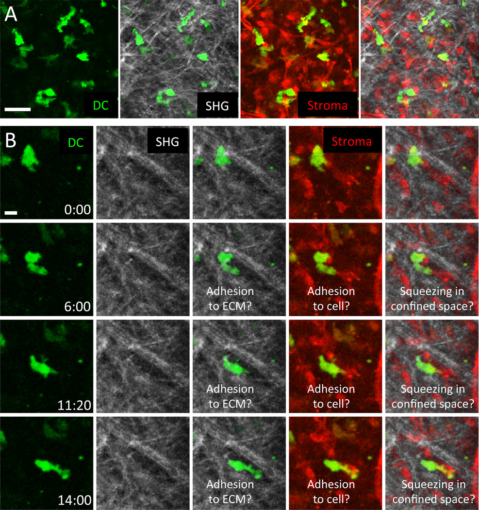
Fig. 4. Optimized analysis of interstitial leukocyte migration in physiological settings.
To dissect cell-intrinsic aspects of leukocyte motility from external guidance, simultaneous imaging of cell dynamics and tissue structures is recommended. (A) 2P-IVM was applied to murine ear skin to visualize the dermis of a transgenic DsRed+/+ CD11c-YFP+/− B6.Albino mouse. Ubiquitously expressed DsRed illuminates all stromal elements (red), YFP-signals highlight endogenous dendritic cells/macrophages (yellow), and SHG signals reflect collagen bundles (white). The mouse was crossed to a B6.Albino background to avoid cell death of light-sensitive melanophages, avoiding laser light-induced artifactual effect in the tissue. Scale bar = 40 µm. (B) The movement of a wild-type dendritic cell, presumably not activated, is followed over 14 min in relation to tissue structures. Simultaneous visualization of cells, stroma, and collagen gives a closer representation of the 3D tissue geometry and reveals channel-like structures and dense networks and surfaces. Still, several other tissue determinants that might influence leukocyte behavior are unknown, e.g., the potential presence of cytokines and chemokines that can induce cell shape changes and upregulation of cell adhesion. Scale bar = 10 µm. CD11c-YFP mice were a kind gift of Michel Nussenzweig.

