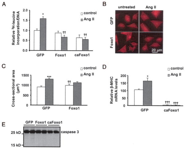
Figure 2.
Foxo1 activity inhibits Ang-II–induced hypertrophic growth in cardiomyocytes. A, Myocytes were cultured and infected with adenovirus and treated with Ang II (48 hours). Cells were then assayed for [3H]leucine incorporation normalized to cellular DNA content. B, Representative fields of cardiomyocytes stained with an α-actinin antibody. C, Quantification of cell cross-sectional area from experiments shown in B. Sixty-five to 75 randomly selected cells from each group were measured. D, β-MHC mRNA levels were measured by real-time PCR. E, Western blots of caspase 3 in whole-cell lysates harvested from myocytes 48 hours after adenovirus infection (A, D, n=8; C, n=65 to 75). *P≤0.05, ***P≤0.001 vs untreated control; ††P≤0.01, †††P≤0.001 vs respective GFP control group.

