
Figure 7. Typical rest cardiac magnetic resonance imaging features in a patient with familial ATTR cardiac amyloidosis.
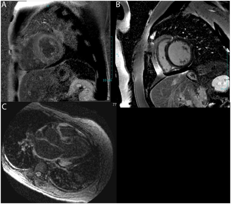
Late gadolinium enhanced images demonstrate diffuse LGE in the left ventricular myocardium (A) contrasted with dark myocardium a normal patient (B). The bottom panel (C) demonstrates LGE in the atrial wall, a characteristic feature of cardiac amyloidosis.