
Figure 2.
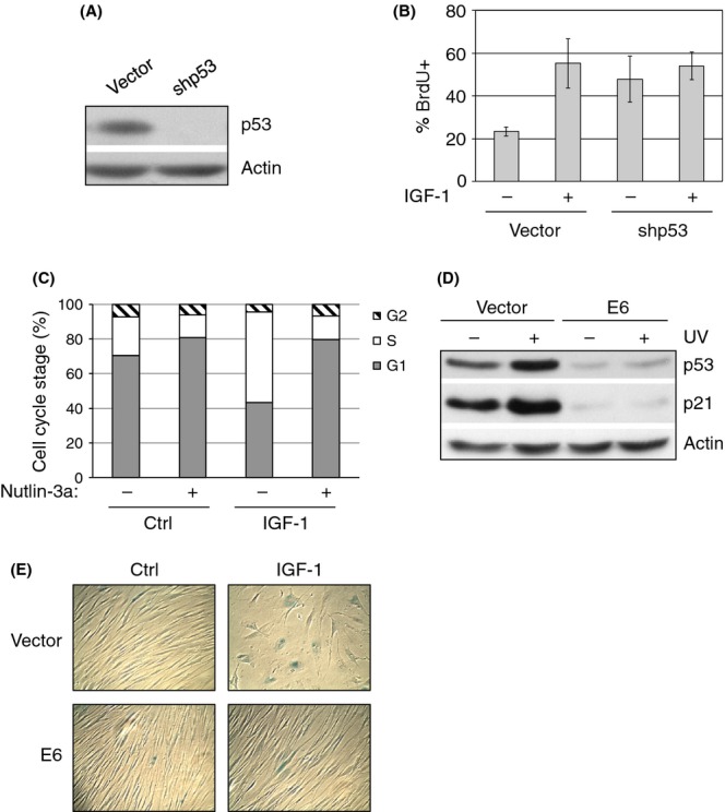
Insulin-like growth factor-1 (IGF-1) induces premature senescence in a p53-dependent manner. (A) MCF7 cells were stably infected with retrovirus expressing shRNA specific for p53 or a vector control. Cell lysates were subjected to western blotting as indicated. (B) MCF7 cells were serum-starved in the absence of FBS for 48 h, then treated with 50 ng mL−1 IGF-1 for 24 h prior to BrdU incorporation assay, as described in the Materials and Methods. BrdU incorporation was quantified as the percentage of BrdU-positive (% BrdU+) cells over the total number of cells. Results are presented as means and SE from three experiments. (C) MCF7 cells were serum-starved for 48 h and then treated with 50 ng mL−1 IGF-1 and/or 5.0 nm Nutlin-3a for 24 h. Cells were then analyzed by flow cytometry. Results are presented as means from two experiments. (D) IMR90 cells stably expressing E6 or vector control were UV irradiated. Cell lysates were subjected to western blot analysis as shown. (E) Stable IMR90 cells were serum-starved for 4 days and then treated with or without IGF-1 for 6 days under serum-starvation conditions and assayed for SA-β-Gal activity. Micrographs are representative of three independent experiments.