
Figure 4.
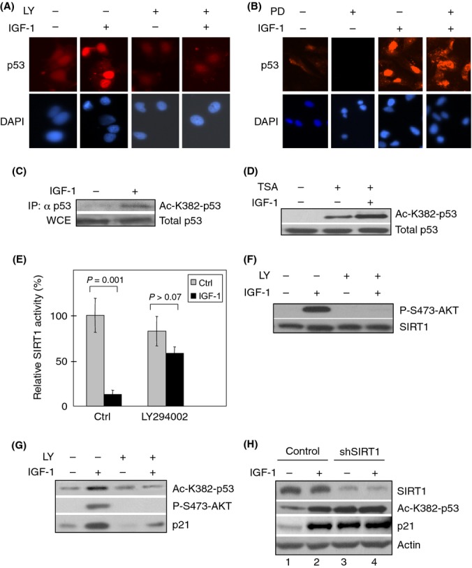
Insulin-like growth factor-1 (IGF-1) inhibits SIRT1 and induces p53 acetylation in a PI3K-dependent manner. (A, B) Serum-starved MCF7 cells were pretreated for 4 h with the PI3K inhibitor LY294002 (LY; 25 μm), the Mek-Erk inhibitor PD98059 (PD; 25 μm) or DMSO as a vehicle control. Cells were then treated with 50 ng mL−1 IGF-1 for 12 h. Cells were subjected to immunostaining for p53 and counterstained with DAPI. (C) Serum-starved MCF7 cells were treated with IGF-1 for 12 h. Whole-cell extract (WCE) were immunoblotted using a pan-specific antibody for p53. Normalized WCE containing comparable amount of total p53 protein were immunoprecipitated and immunoblotted with an antibody specific for acetylated p53 (Ac-K382-p53). (D) MCF7 cells were serum-starved for 48 h and treated with IGF-1 for 12 h. Cells were then treated with 40 μm Trichostatin A (TSA) for 6 h before collection. Whole-cell extracts were subjected to western blotting as indicated. (E–G) Subconfluent MCF7 cells were serum-starved for 48 h and pretreated for 2 h with 25 μm LY294002 or vehicle (Ctrl), prior to treatment with IGF-1 for 12 h. (E) Equal amounts of whole-cell extracts were subjected to SIRT1 immunoprecipitation for in vitro Fluor de Lys (Biomol) deacetylation assay. Relative SIRT1 activity is presented as means and SE from two independent experiments performed in duplicate. (F) Western blot analyses were performed to ensure comparable SIRT1 protein input from IP for Fluor de Lys assay and for analysis of IGF-1-induced AKT activation. (G) Whole-cell extracts were subjected to western blot analysis as shown. (H) MCF7 cells were stably infected with retrovirus expressing shRNA specific for SIRT1 or a control and selected by puromycin resistance. Cells were serum-starved for 48 h and treated with 50 ng mL−1 IGF-1 for 12 h prior to treating with 40 μm Trichostatin A for 6 h. Whole-cell extracts were subjected to western blotting.