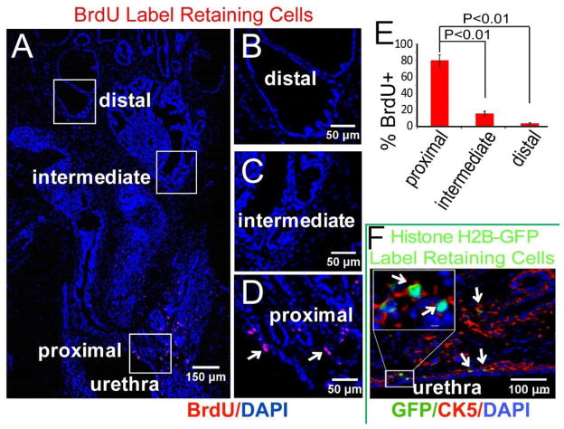
Figure 1. Location of label retaining cells in the adult mouse prostate.
(A) Wild-type CD-1 dams were injected with BrdU at E16 and the male offspring harvested at 10 weeks. Immunofluorescence staining localized BrdU label-retaining cells mostly to the proximal duct segments and junction with the urethra. (Montage image of sagittal sections of prostate). (B–D) Boxed regions in image A. (E) Distribution of LRCs in proximal, intermediate and distal regions (n=5). (F) H2B-GFP mice were mated and GFP expression from the cytokeratin 5 (CK5) promoter was activated in pregnant females for 24 hours at E16 by removing doxycycline from the water supply. The male offspring were harvested at 10 weeks, and fluorescence microscopy localized most GFP label-retaining cells to CK5 (red) positive cells.

