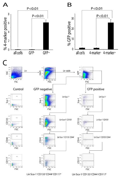
Figure 2. GFP labeling at E16 yields a population of labeled cells enriched for co-expression of stem cell markers.
CK5/H2B-GFP mice were GFP-labeled at E16 and sacrificed at 10 weeks. Single cells were isolated by enzymatic digestion and lineage committed cells were depleted for Lin− cells. FACS analysis of Lin− was performed for co-expression of Sca-1, CD133, CD44 and CD117 (n=3). (A) Approximately 6 % of GFP positive cells co-expressed these 4 markers, while only 0.1% of the total cell populations co-expressed these 4 markers. (B) Approximately 26% of cells co-expressing these 4 markers were GFP positive, while only 1.5% of the total cell population was GFP positive. (C) Diagram of sequential analysis of flow cytometry data. Cells from the prostate were isolated by magnetic beads to obtain Lin− cells. These cells were gated by forward scatter (FSC) and side scatter (SSC). Then the cells were gated by DAPI exclusion (Lin− viable). Lin− viable cells were gated by GFP expression to separate GFP+ and GFP− cells. GFP+ and GFP− cells were analyzed by sequential gating for Sca-l, CD133, CD44 and CD117 and the percentage of 4-marker positive cells in GFP+ and GFP− groups were determined.

