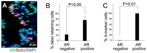
Figure 3. The majority of prostate 4-marker cells stain positive for androgen receptor.

CD-1 mice were BrdU labeled at E16 and sacrificed at 10 weeks. Co-staining was performed for androgen receptor (AR) and BrdU. (A) A representative section of co-stained prostate epithelium. (B) Quantitative analysis of co-staining for AR and BrdU in the prostate epithelium (n=5). (C) Ten weeks old CD-1 mice were sacrificed and the epithelium subject to FACS analysis for AR, Sca-1, CD133, CD44 and CD117. Quantitative analysis shows that most 4-marker cells are androgen receptor positive (n=3).
