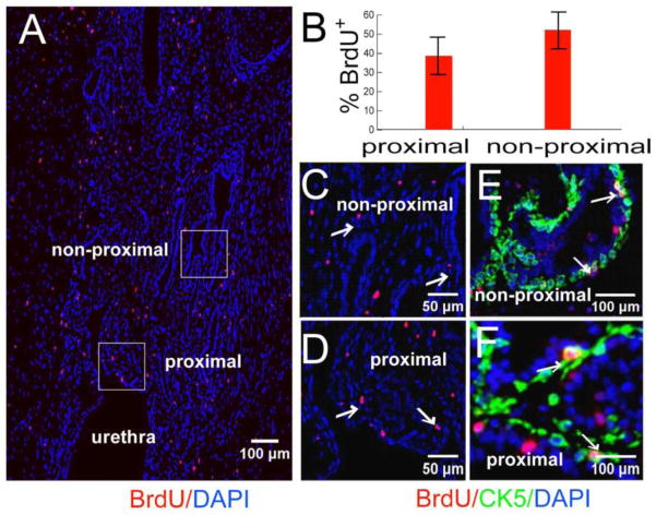
Figure 4. BrdU labeled cells are dispersed post-castration.
CD-1 mice BrdU labeled at E16 were castrated at 8 weeks and sacrificed 2 weeks after castration. (A) Immunostaining for BrdU labeled cells (Montage image of sagittal sections of prostate). (B) Quantitative analysis for the location of BrdU labeled cells (n=5). (C and D) Boxed regions in A. (E and F) Colocalization of BrdU label-retaining cells (red) with CK5 (green) in non-proximal and proximal ducts.

