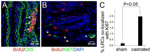
Figure 5. Castration induces proliferation of BrdU labeled cells.

(A) Clusters of BrdU labeled cells (arrows) were observed in mice labeled and castrated as described in Figure 4. Similar clusters were never observed in the intact controls. Co-staining for BrdU and CK5 identified the cluster of labeled cells as basal cells. (B and C) Staining for Ki67 in mice labeled and castrated as described in Figure 4 revealed increased proliferation of BrdU labeled cells. (B) Immunofluorescence staining for BrdU (red), Ki67 (green) and DAPI (blue) shows co-localization of BrdU with Ki67. This is quite uncommon in intact controls. Quantitative analysis of co-staining for Ki67 and BrdU confirms co-staining is significantly increased castrated animals (C) (n=5).
