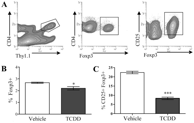
FIGURE 1.
The frequency of Foxp3+ donor CD4+ cells is decreased in mice exposed to TCDD during acute GVH response. A, Using flow cytometry, B6 donor CD4+ cells were identified in vehicle- or TCDD-treated F1 host mice at 48 h after adoptive transfer by their expression of the congenic marker Thy 1.1, from which Foxp3, and/or CD25 were measured. B and C, The percentage of Thy1.1+ CD4+ cells (B) and percentage of Thy1.1+CD4+ CD25+ cells (C) expressing Foxp3 is shown. These data are representative of two separate experiments (n = 3 mice per treatment group). Asterisks indicate statistically significant difference in the percent of Foxp3+ cells compared with vehicle control (t test; *, p < 0.05; ***, p < 0.0005).

