
Fig. 4.
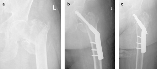
AP X-ray views of left proximal Femur demonstrating (a) an unstable (31A2) intertrochanteric fracture treated with (b) a DHS, restoring proximal femur anatomy, but with (c) shortening of femoral neck and reduced femoral offset at union.
Official websites use .gov
A
.gov website belongs to an official
government organization in the United States.
Secure .gov websites use HTTPS
A lock (
) or https:// means you've safely
connected to the .gov website. Share sensitive
information only on official, secure websites.
AP X-ray views of left proximal Femur demonstrating (a) an unstable (31A2) intertrochanteric fracture treated with (b) a DHS, restoring proximal femur anatomy, but with (c) shortening of femoral neck and reduced femoral offset at union.