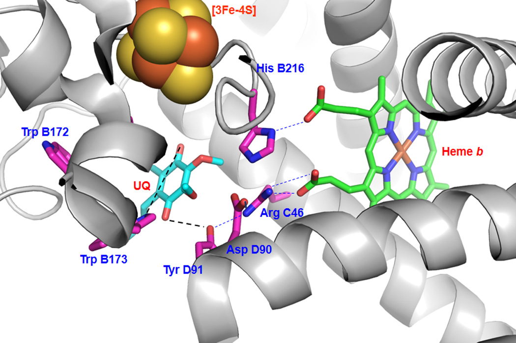
Figure 3.

Hydrophobic residues and polar interactions (dashed lines) in the ubiquinone-binding site (Qp site) of mammalian complex II. The quinone-binding pocket is revealed by the X-ray structure (pdb 1ZOY), and involves the residues Trp B172, Trp B173, His B216, Arg C46, Asp D90, Tyr D91. The residues of the ubiquinone-binding site are determined by X-ray structure to be Trp B173 and Tyr D91. UQ denotes ubiquinone.
