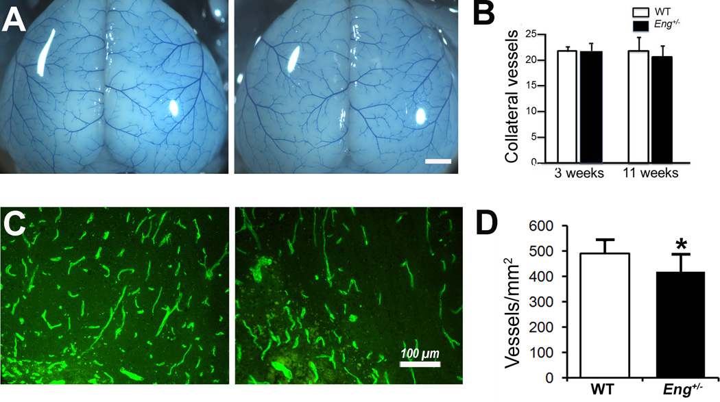
Figure 4. Pial collateral vessels at baseline and vessel density in the peri-infarct cortex.
A. Representative photographs of collateral vessels of 3-week-old WT and Eng+/− mice. Bar=1 mm. B. Quantification of collateral vessel number of 3-week-old mice (WT: N=6, Eng+/−: N=6) and 11-week-old mice (WT: N=8, Eng+/−: N=4). C. Representative images of CD31 antibody-stained sections. Bar=100µm. D. Quantification of vessel density. *P=0.05, N=6.

