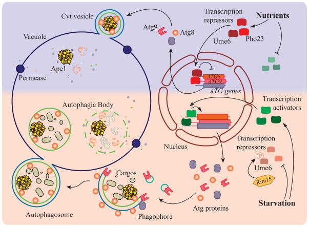
Figure 1.
A model of transcriptional regulation of autophagy in yeast. When nutrients are present, the transcription of most ATG genes is repressed due to the inhibition of autophagy transcription activators, and/or activation of transcription repressors. For example, Ume6 represses the transcription of ATG8, and Pho23 represses the expression of ATG9. Repression of transcription in growing conditions leads to a relatively low amount of Atg proteins that are sufficient for basal autophagy, and consequently the autophagosomes that are formed under these conditions are predicted to be smaller in size (due to limited Atg8) and generated at a slower rate (due to limited Atg9). The Cvt pathway, which is the main type of autophagy-like process in growing conditions, generates Cvt vesicles to sequester prApe1 dodecamers and deliver them to the vacuole to allow maturation of the zymogen. After starvation, transcription activators of autophagy become functional, and the repressors are inhibited. For example, Ume6 is phosphorylated by Rim15 kinase, releasing its repression of ATG8 transcription. Upregulation of ATG gene expression allows larger amounts of Atg components, such as Atg8 and Atg9, to participate in the formation of the autophagosome. As a result, more and larger autophagosomes are generated. During starvation-induced autophagy, Atg proteins are initially recruited to a peri-vacuolar site to generate the double-membrane structure named the phagophore. The phagophore randomly sequesters cytoplasmic material, and after the expansion phase is complete it seals to generate the double-membrane autophagosome. The outer membrane of the autophagosome fuses with vacuole, releasing the inner vesicle, now termed an autophagic body. This single-membrane vesicle along with its cargo is degraded in the vacuole lumen, and the resulting macromolecules are released back into the cytosol.

