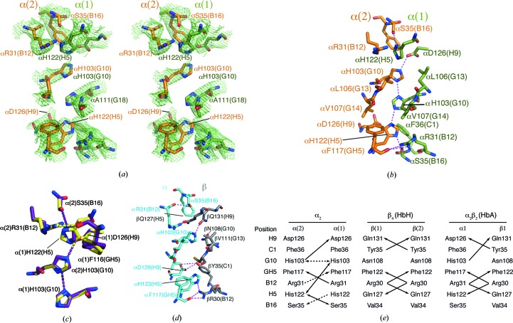
Figure 2.
The α2 dimer interface. (a) The OMIT map electron density of the α2 dimer interface contoured at 1σ (mesh), showing the four buried His side chains. The OMIT map was calculated using the SFCHECK program from CCP4 (Winn et al., 2011 ▶). (b) Hydrogen-bonding interactions at the α2 dimer interface (PDB entry 3s48). (c) Comparison of interfacial residues and hydrogen-bonding interactions in PDB entry 3s48 (violet) and the structure re-refined by PDB_REDO (yellow). (d) Hydrogen-bonding interactions at the α1β1 interface of HbA (PDB entry 2dn3; Park et al., 2006 ▶). (e) A comparison of the hydrogen-bonding interactions at the α1β1-equivalent interfaces of α2, β4 and α2β2. Double-headed arrows indicate hydrogen bonds where the donor/acceptor atoms could potentially be reversed, for example owing to protonation of α(2)His103 under the influence of α(1)Asp126. Dashed lines indicate hydrogen bonds that appear in 3s48 or the PDB_REDO re-refinement of 3s48 but not both.

