Figure 3.
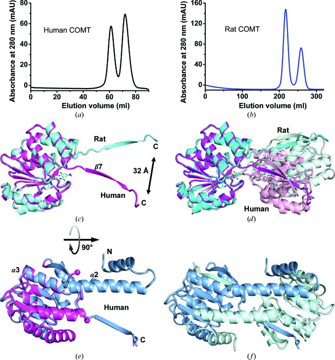
Domain swapping in COMT. (a) Gel-permeation chromatography of human COMT on a Superdex S75 16/60 column shows peaks for monomeric and dimeric COMT. Human COMT was placed into the same buffer as rat COMT (see below), or sometimes in 50 mM HEPES–NaOH pH 7.0, 150 mM NaCl, 2 mM MgCl2, 1 mM TCEP, 10% glycerol. The buffer has no noticeable influence on the monomer:dimer ratio. (b) The same experiment for rat COMT on a larger column (home-made S75 26/80), resulting in better separation of the species. The buffer was 50 mM Tris–HCl pH 7.5, 50 mM NaCl, 10 mM DTT, 2 mM MgCl2. The methyltransferase activities do not differ appreciably between monomer and dimer preparations (data not shown). (c) Superposition of rat (cyan; PDB entry 3oe4) and human [magenta; structure (2)] structures that display a swap of their C-terminal β7 strand. The bisubstrate inhibitor in 3oe4 is drawn as a stick model. Its nitrocatechol part would clash with the conformation of the human apo COMT structure, offering an explanation for the discrepancy of the β7 vectors. (d) The resulting dimers (lighter hues) therefore show a very different juxtaposition of the monomers when rat and human COMT are compared. (e) Example of a double domain swap of human apo COMT (3). In addition to the C-terminal β7 strand, the first 40 residues encompassing helices α1 and α2 have swapped [light blue; structure (3)]. Structure (2) is superimposed as a reference. The disordered α2/α3 loop between Lys86 and Gly93 in structure (2) (human numbering) is marked with spheres. This loop forms part of the α2 helix in structure (3), where these residue positions are also marked. The view in (e) is rotated 90° compared with that in (c). (f) The dimer formed by the double domain swap.

