
Figure 4.
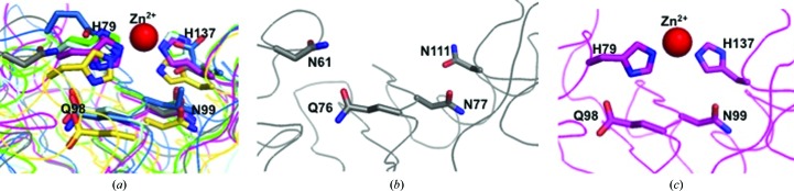
The CAP cavity. (a) The superposed central cavity of CAPs reveals that key residues corresponding to the Zn2+-binding site superimpose well in representative CAP structures. CAP structures are colored as follows: SmVAL4, gray; NaASP2, green; tablysin-15, blue; GAPR-1, yellow; sGLIPR1, magenta; vCRISP, cyan. The numbers correspond to those for GLIPR1 and the Zn2+ ion is shown as a red sphere. (b) The same region and view for SmVAL4 alone reveals the absence of the His that coordinates Zn2+; numbering corresponds to that of SmVAL4. (c) The same region and view for sGLIPR1 alone; numbering corresponds to that of sGLIPR1.