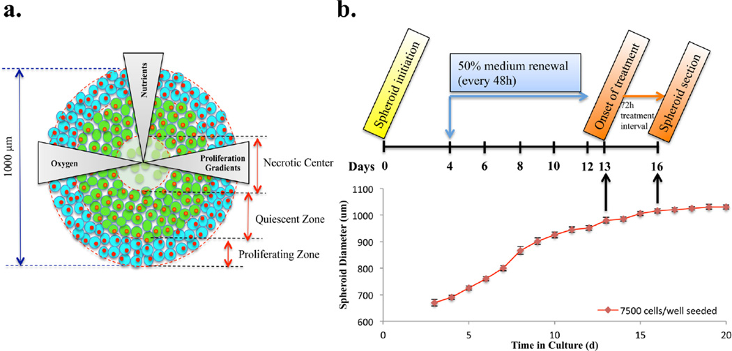
Figure 1.
Timetable for setting up spheroid-based drug evaluation. (a) Cartoon structure of the in vitro tumor spheroid. (b) Schematic illustration of dosing procedures and diameter growth of HCT 116 spheroids as a function of time. Photographs (n = 8) were acquired every day and spheroid diameters were calculated using a hemocytometer scale bar. The interwell variation in spheroid diameter was under 5%.

