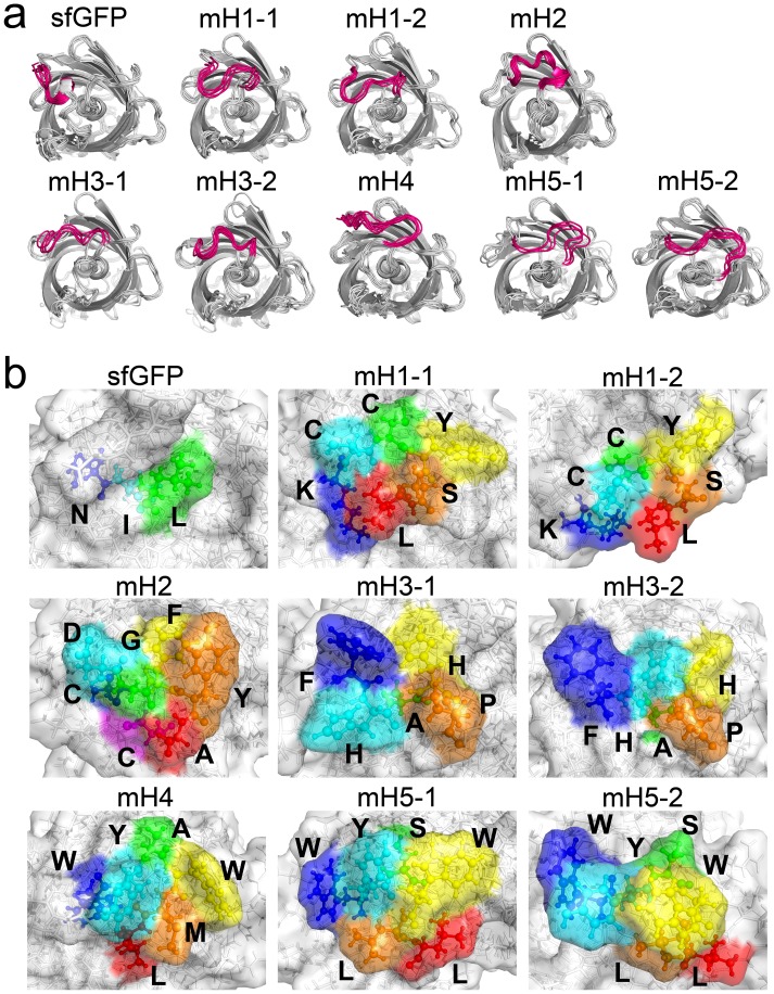
Figure 4. Analysis of the gFPSs containing HER2-BPs.
(a) Superimposed structures of sfGFP, mH1, mH2, mH3, mH4, and mH5 at every 0.5 ns throughout the 4.5–9.0 ns trajectory of the MD simulations. The K131–L137 integration sites are highlighted in magenta. (b) Representative surface structures for sfGFP, mH1, mH2, mH3, mH4, and mH5. Amino acids (N135–L137) in the sfGFP and integrated HER2-BPs of mH1–mH5 are also shown using the ball and stick model. These peptides are highlighted in blue, cyan, green, yellow, orange, red, and magenta for the 1st–7th amino acids, respectively.

