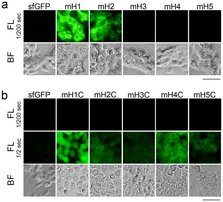
Figure 5. Binding assays for the gFPSs containing HER2-BPs.
(a) Fluorescence (FL) and bright field (BF) micrographs of HER2-positive N87 cells treated with sfGFP, mH1, mH2, mH3, mH4, and mH5 for 16 h. Exposure time = 1/200 s. Bar = 50 µm. (b) FL and BF micrographs of HER2-positive N87 cells treated with sfGFP, mH1C, mH2C, mH3C, mH4C, and mH5C for 16 h. Exposure time = 1/200 s or 1/2 s. Bar = 50 µm.

