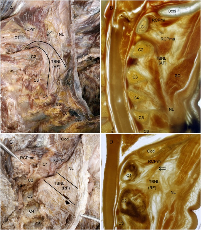
Figure 1. The to be named ligaments were shown in the dissected specimens and the P45 plastination sheets.
A, C: Lateroposterior aspect of the nuchal ligament; B, D: Median sagittal section of the head-neck in a P45 plastination sheet. To Be Named Ligament (TBNL) within the nuchal ligament was formed by arcuate fibers (A, B). The arcuate fibers arose from the lower part of the posterior border of NL below the level of the spinal process of vertebra C3 (A, B), continuing anterosuperiorly crossing over the spinous process of axis and continuing into the atlantoaxial interspace (B). Its path continues traversing through the interspace and attaching to the posterior aspect of the cervical dura mater (B). The rectus capitis posterior minor (RCPmi) emitted a bundle of muscular fibers (↓) which were attached to TBNL (A). Another kind of TBNL within the nuchal ligament was formed by radiated fibers (C, D). The radiated fibers originated from the posterior border of the upper part of nuchal ligament opposite to the spinal processes of vertebrae C2 and C3. At this point the radiated fibers ran anteriorly to traverse through the atlantoaxial interspace and finally attached to the posterior aspect of the cervical dura mater (D). Additionally, the RCPmi emitted a bundle of muscular fibers (↓↓), which merged into TBNL before it enterd the atlantoaxial interspace (D). TBNL, To Be Named Ligament. NL, nuchal ligament. AF, arcuate fibers of TBNL. RF, radial fibers of TBNL. C1∼C7, first to seventh cervical vertebra; Occi, occipital bone; RCPma, rectus capitis posterior major; SC, splenus capitis.

