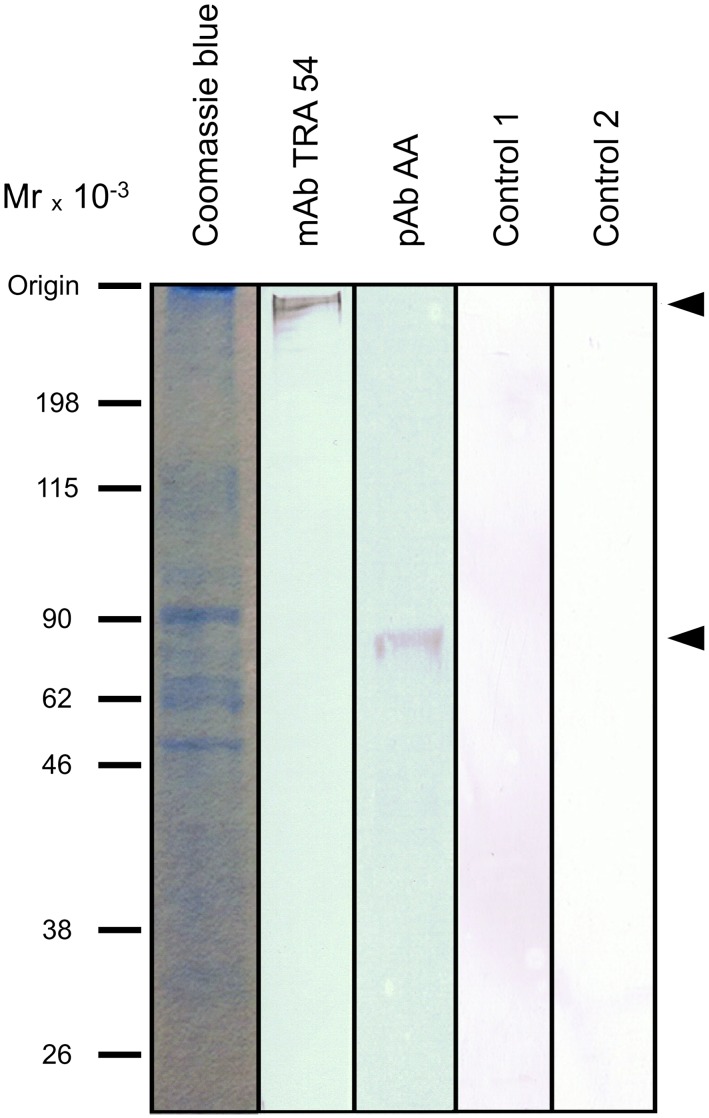
Figure 1. Immunoblots of mouse caput epididymis extracts.
Aliquots (50 µg) of protein were electrophoresed in a 10% polyacrylamide gel and transferred to a PVDF membrane for immunoblotting with mAb TRA 54 or pAb rabbit anti-albumin (Abcam, Tokyo, Japan, ab34807). Arrows indicate the upper (mAb TRA 54) and lower (pAb anti-albumin) limits of the protein complexes detected in the samples. Control 1 is an immunoblot without mAb TRA 54 and control 2 is a kidney extract immunoblotted with mAb TRA 54. No immunoreactive bands were detected in either of these controls. Molecular mass markers (MM) are shown on the left.

