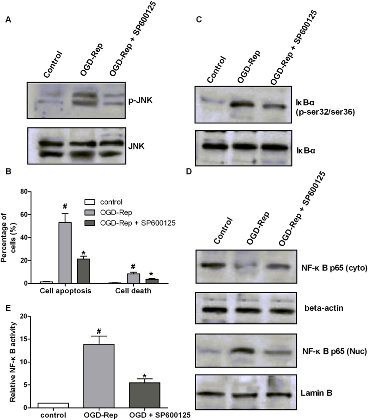
Figure 8. Inhibition of JNK phosphorylation phenocopies the effect of ginsenoside Rb3.
H9c2 cells were treated with a JNK inhibitor, SP600125, followed by OGD-Rep. (A) Cell lysates were collected and subjected to Western blot assay to analyze the JNK phosphorylation and JNK protein levels. (B) The cell apoptosis was determined by Annexin V-PE/7-AAD assay. The graph represented the percentage of apoptotic and dead cells. SP600125 decreased the cell apoptosis and cell death that was induced by OGD-Rep. (C) The cell lysates were collected and subjected to Western blot assay to analyze the phosphorylation of IκB-α and total IκB-α protein. SP600125 decreased the IκB-α phosphorylation at ser32/ser36 sites that was induced by OGD-Rep. (D) The nuclear and cytoplasmic proteins were isolated and sunjected to Western blot assay to analyze the protein levels of p65 in the nucleus and cytoplasma. Lamin B was used as an internal control to normalize the nuclear p65, and beta-actin was used as an internal control to normalize the cytoplasmic p65. SP600125 decreased nuclear p65 levels and increased cytoplasmic p65 levels compared to the cells with OGD-Rep. (E) The NF-κB activity was analyzed using a NF-κB p65 ELISA Kit, and the graph represented the ratio of the nuclear to cytoplasmic p65 concentration. All data were shown as the mean ± SD. #P<0.05 vs. group without OGD-Rep. *P<0.05 vs. group treated with OGD-Rep alone. n = 3–6.

