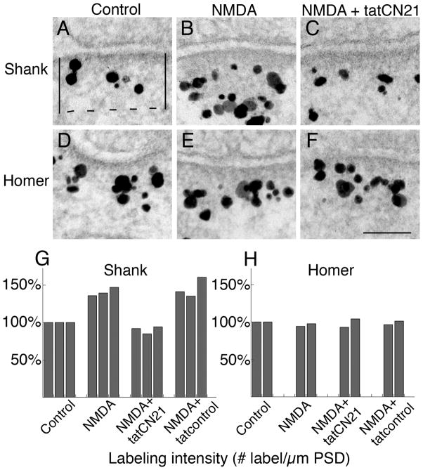
Figure 1. NMDA-induced Shank increase at the PSD is dependent on CaMKII activity.
Hippocampal neurons in culture were pre-incubated for 20 min with or without tat-peptides, as indicated, and then were treated with NMDA-containing media with the same additions or control medium for another 2 min. The cells were then immediately fixed and prepared for antibody labeling.
Above Electron micrographs of synapses labeled for Shank (A–C) or Homer (D–F). Gold labels appear as black particles of heterogeneous size. Label for Shank increased at the PSD complex after NMDA treatment (B vs. A), and this increase was blocked by the CaMKII inhibitor, tatCN21 (C). In contrast, distribution of label for Homer remained unchanged under these same conditions.
Below bar graphs. PSD measurement area was marked as shown in (A) and labeling intensity was calculated as number of labels per μm PSD length. Labeling intensity at the PSD complexes was normalized to control values for Shank (G, three experiments with a pan Shank antibody) and Homer (H, one experiment with a pan Homer1 antibody and a second experiment with a Homer 1b/c antibody). Statistical analyses were carried out via ANOVA with Tukey’s post test within each experiment:
For Shank, between control and NMDA, P< 0.005 for exp 1, P<0.001 for exp 2, P<0.01 for exp 3; between NMDA and NMDA+tatCN21, P<0.0001 for all 3 exp; between NMDA+tatCN21 and NMDA+tatcontrol, P<0.0001 for all 3 exp. For Homer, there were no statistically significant differences among any two treatment conditions.

