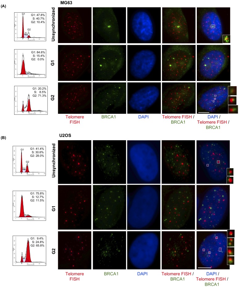
Figure 1. BRCA1 localizes to ALT cell telomeres primarily in G2 of the cell cycle.
(A) MG63 and (B) U2OS cells were synchronized by double thymidine block. Asynchronous and synchronized cells were collected and analyzed by flow cytometry to determine cell cycle distribution. Cells were fixed at corresponding phases of cell cycle and stained with antibodies to BRCA1 (green); telomeres were labeled by FISH with a PNA probe (red); nuclei were stained with DAPI (blue). Images shown are a single maximum intensity projection (MIP) image. Enlargements of co-localizations are shown at the right. Scale bar: 10 µm.

