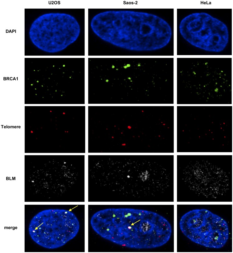
Figure 3. BRCA1 and BLM co-localize at telomeres in ALT cells.
(A) Cells were synchronized to G2 (7h post release after double thymidine block) and stained with antibodies to BRCA1 and BLM and telomeres labeled by FISH with a PNA probe (upper panel) as in Figure 1. Yellow arrows indicate foci with BLM-BRCA1-telomere co-localization.

