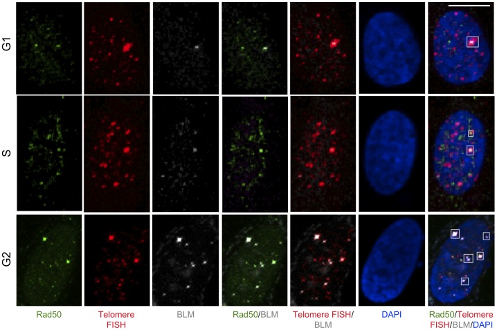
Figure 7. Co-localization of BLM and RAD50 at telomeres at different stages of cell cycle in ALT cells.
U2OS cells were synchronized by double thymidine block in G1-, S- and G2-stages of the cell cycle. Cells were fixed and stained with antibodies to BLM (grey), Rad50 (green) and telomeres labeled by FISH with a PNA probe (red). Nuclei are stained with DAPI. Images are shown as MIP image. White boxes indicate co-localizations of BLM, Rad50 and telomere FISH. Scale bar: 10 µm.

