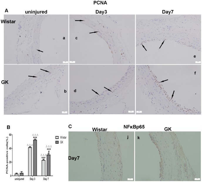
Figure 4. Immunohistochemistry for the expression of PCNA and NFκBp65 in common carotid arteries.
PCNA-positive cells of uninjured in both rats (A–a, A–b). PCNA-positive cells of Day 3 post-injury (A–c, A–d) were more than those of Day 7 post-injury (A–e, A–f) in both rats and PCNA-positive cells in GK rats were more than those in Wistar rats (B). NFκBp65-positive cells (C) in GK rats were more than those in Wistar rats at Day 7 post-injury. Data are represented as mean ± SEM. ΔΔΔindicates versus uninjured, p<0.001; ***indicates versus Day 3 post-injury, p<0.001; ##indicates versus Day 7 post-injury, p<0.01.

