
Figure 6. Histological studies in the cerebellum and skeletal muscle.
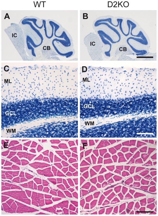
Microphotographs showing representative images of Nissl-stained tissue sections from cerebellum (A–D) and hematoxylin-eosin stained tissue sections from gastrocnemius skeletal muscle (E,F) in 6-month-old WT (A, C, E) and D2KO (B, D, F) mice. A, B Low magnification images showing a complete cerebellum in a tissue section. C, D Detail of cerebellar layers. Gross structural alterations were found neither in cerebellum (A–D) nor in skeletal muscle (E,F). Tissues from 3 animals of each experimental condition were used in these studies. CB: cerebellum, ML: Molecular layer of the cerebellum, GCL: granule cell layer of the cerebellum, WM: white matter, IF: inferior colliculus. Scale bars, 1 mm for A–B and 100 µm for C–F.