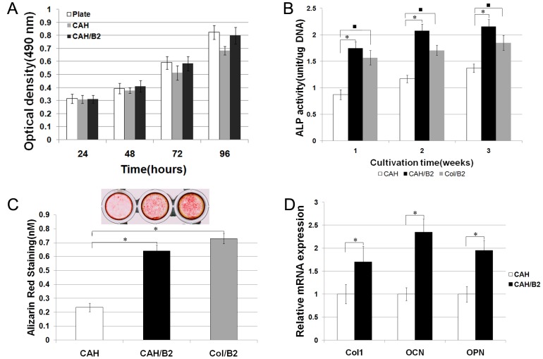
Figure 3. Biocompatibility and effects of the CAH/B2 scaffold on osteoblastic differentiation.
A. MTS assay of MSCs cultured with CAH and CAH/B2 scaffolds. The cells were also cultured in plates as a control. Data represent the mean+SD of n = 5 samples. No statistically significant differences were seen between groups. B. Effects of CAH/B2 on in vitro ALP activity. ALP activity in the CAH/B2 group was higher than in the CAH group. One-way analyses of variance suggest that there are significant differences among three groups. The significant post hoc test results are identified by symbol *. N = 5. P<0.001: CAH vs. CAH/B2. C. The level of calcium deposition in 3-week cultures was evaluated by AR-S. The values indicated are means ± SD, (n = 5) p<0.005 as compared with that deposition in the CAH group. D. qRT-PCR analysis of osteoblast marker genes, showing that MSCs cultured on a CAH/B2 scaffold exhibited increased Col I, OCN and OPN gene expression. Data represent the mean + SD of n = 5 samples. P<0.05.

