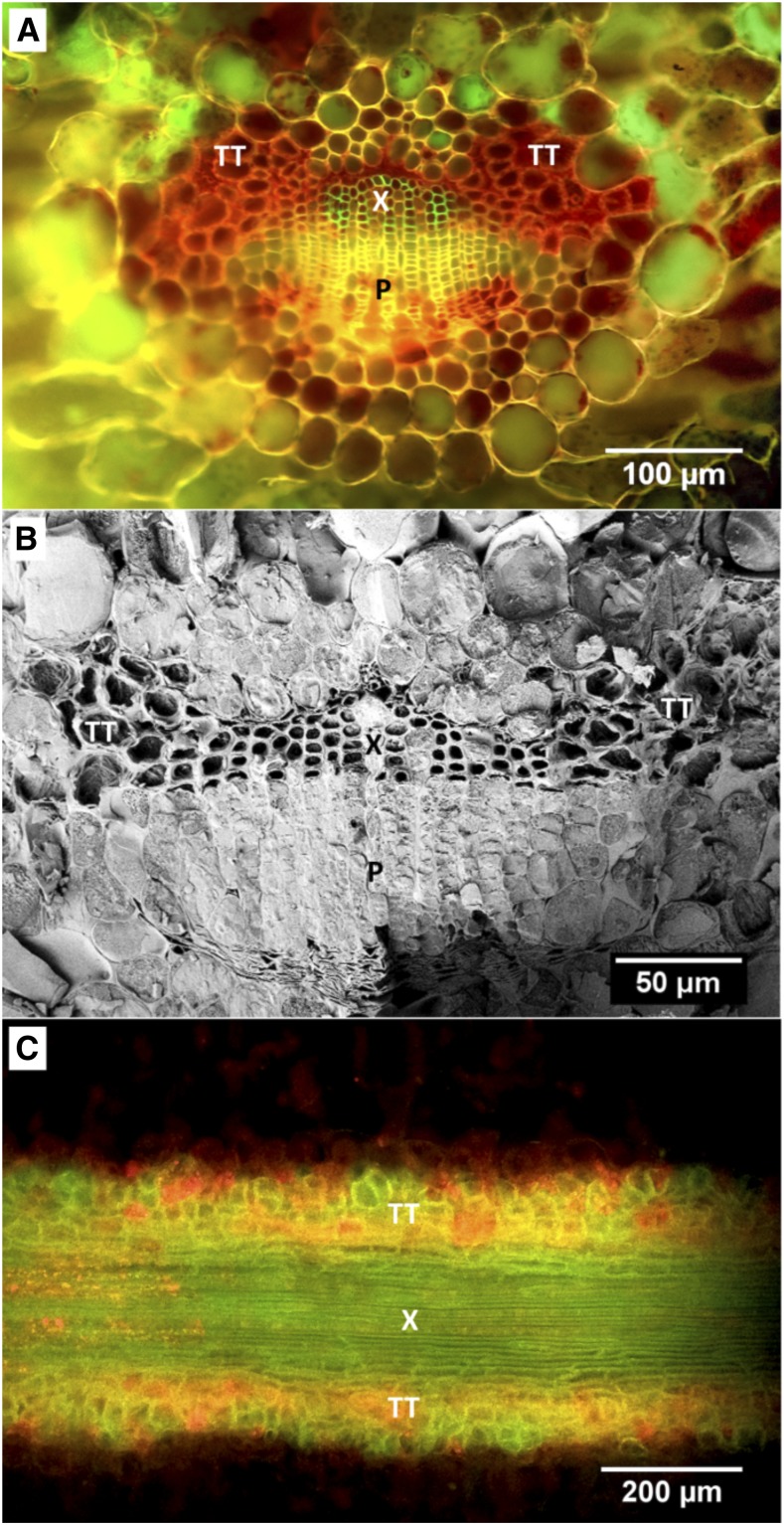
Figure 1.
Structure and position of transfusion tissue in relation to the axial vascular system in leaves of T. baccata. A, Cryo-fluorescence image of a cross section of a T. baccata leaf after infusion with acid fuchsin. Tissues are xylem (X), phloem (P), and transfusion tracheids (TT). B, CSEM image of a cross section of a hydrated T. baccata leaf. The sample was heavily etched to sublimate xylem and transfusion tracheid sap, which does not have high concentrations of solutes, to show the cell wall structure of xylem and transfusion tracheids. C, Fluorescence image of a paradermal section of the leaf midvein stained with phloroglucinol/HCl.

