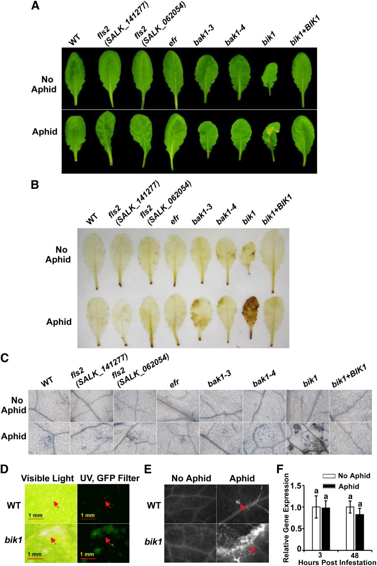
Figure 2.
Aphid infestation induces a heightened HR in bik1. Representative leaf images of lesion formation (A), DAB staining (B; H2O2 indicator), and trypan blue staining (C; cell death indicator) prior to (top) or 6 d after (bottom) aphid infestation of genotypes indicated. D, Autofluorescence of aphid-induced lesion spots under UV excitation with GFP filter set (right). The same fields of view are shown under visible light (left). E, Callose deposition at lesion sites. Left, control leaves; right, callose deposition after aphid treatment. Arrows point to lesion sites. F, Relative expression of BIK1 in wild-type (WT) plants in the presence and absence of aphid infestation. Three-week-old plants were infested with aphids as described in “Materials and Methods.” Data were analyzed by independent samples’ Student’s t test. Means with different letters were significantly different (P < 0.05).

