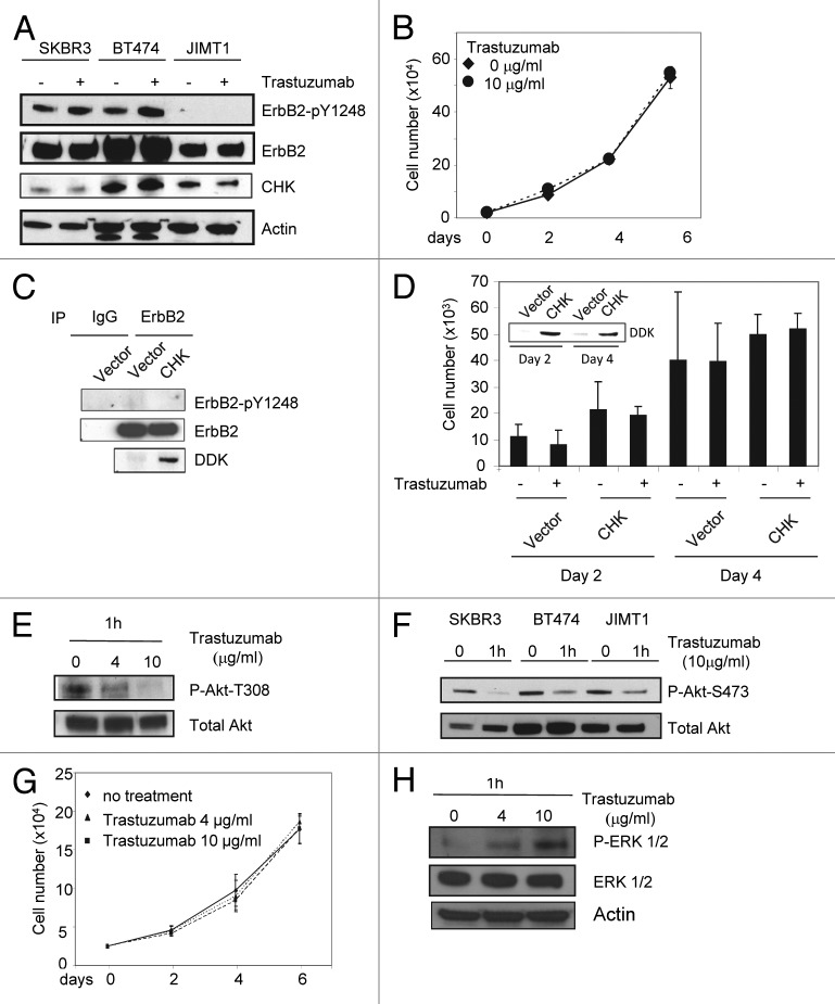
Figure 6. Trastuzumab is unable to induce ErbB2-Y1248 phosphorylation in JIMT1 cells, but is still capable of inhibiting Akt activity. (A) Analysis of ErbB2-Y1248 phosphorylation in trastuzumab-sensitive and trastuzumab-resistant cells as indicated following treatment with trastuzumab. Cells were plated in 10% serum containing media for 24 h and then treated with trastuzumab (4 μg/mL) for 1 h or left untreated. The levels of ErbB2-pY1248, total ErbB2, and endogenous CHK in WCL were detected by western blot analysis using anti-phospho-ErbB2-Y1248, anti-ErbB2, and anti-CHK antibodies. (B) JIMT1 cells were plated at 2.5 × 10⁴/well in 12-well plates in triplicate in media containing either trastuzumab (10 μg/mL) or no trastuzumab (control). Cells were trypsinized, mixed with trypan blue, and counted at the indicated times using a BioRad TC10 automated cell counter. Data represent the mean ± SEM from one of the two independent experiments, and each was performed in triplicate. (C) JIMT1 cells were electroporated with either empty pCMV6-entry vector or pCMV-entry vector encoding DDK-tagged CHK. WCL were harvested 48 h post-transfection and then subjected to immunoprecipitation using an antibody directed against ErbB2. The levels of ErbB2-pY1248 in immuneprecipitates were evaluated by western blot analysis using an antibody directed against ErbB2-pY1248. Total ErbB2 in the immunoprecipitates was detected using an antibody directed against ErbB2. The levels of DDK-CHK in WCL were detected using an antibody directed against DDK tag. IgG was used as a negative control. (D) JIMT1 cells were electroporated with either empty pCMV6-entry vector or pCMV-entry vector encoding DDK-tagged CHK. Cells then were seeded at 10 × 103/well in 12-well plate in triplicate and treated with trastuzumab (50 μg/mL) or left untreated. Cells were trypsinized, mixed with trypan blue and counted using a BioRad TC10 automated cell counter on the indicated days. Data represent the mean ± SEM from one of the two independent experiments, and each was performed in triplicate. Inset: Levels of DDK-CHK expression in WCL were detected in JIMT1cells by western blot analysis using an antibody directed against DDK at the indicated times. (E) JIMT1 cells were plated in media containing 10% serum for 24 h and then serum-starved for another 24 h. Cells then were treated with trastuzumab at the indicated concentration for 1 h or left untreated. The levels of P-Akt-T308 and total Akt in WCL were detected by western blot analysis. (F) The experimental procedures were essentially the same as described in (E) except that SKBR3 and BT474 cells were also included and the levels of P-Akt-S473 and total Akt in WCL were detected by western blot analysis. (G) MCF7 cells were plated at 2.5 × 10⁴/well in a 12-well plate overnight, and then treated with either trastuzumab (4 and 10 μg/mL) or left untreated for the indicated days. Cells were trypsinized, mixed with trypan blue, and counted using BioRad TC10 automated cell counter on the indicated days. Data represent the mean ± SEM from one of the two independent experiments, and each was performed in triplicate. (H) P-ERK1/2 and total ERK1/2 detected in MCF7 WCL by western blot analysis using antibodies directed against P-ERK1/2 and ERK1/2, respectively.

An official website of the United States government
Here's how you know
Official websites use .gov
A
.gov website belongs to an official
government organization in the United States.
Secure .gov websites use HTTPS
A lock (
) or https:// means you've safely
connected to the .gov website. Share sensitive
information only on official, secure websites.
