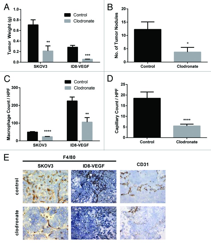
Figure 1. Clodronate treatment reduces tumor burden, TAM levels, and tumor capillary density. (A) Quantification of tumor weight (g) harvested from female mouse SKOV3ip1 and ID8-VEGF ovarian tumor models treated with 0.2 or 1.0 mg/mouse clodronate-encapsulated liposomes, respectively (n = 4 [SKOV3ip1] and 4 [ID8-VEGF] mice) or equally diluted control liposomes (n = 8 [SKOV3ip1] and 6 [ID8-VEGF] mice) 1 d after last injection showed significantly decreased tumor weight in the clodronate treatment group relative to the control group. **P < 0.01 and ***P < 0.0009 determined by the Student t test. Data are shown as means ± the standard error of the mean (SEM). (B) Quantification of tumor nodules present in the peritoneal cavity of female nude mice in the SKOV3ip1 ovarian tumor model group treated with 0.2 mg/mouse clodronate-encapsulated liposomes (n = 4) or equally diluted control liposomes (n = 8) 1 d after last injection showed significantly fewer number of tumor nodules in the clodronate treatment group relative to the control group. *P < 0.03 determined by the Student t test. Data are shown as means ± SEM. (C) Quantification of F4/80 stained tumor sections taken from female mouse SKOV3ip1 and ID8-VEGF ovarian tumor models treated with 0.2 or 1.0 mg/mouse clodronate-encapsulated liposomes or equally diluted control liposomes showed significantly decreased macrophage density in the clodronate treatment group relative to the control group. n = 5 mice for the SKOV3ip1 group and n = 3 mice for the ID8-VEGF group. ****P < 0.0001 and **P = 0.0014 as determined by the Student t test. Data are shown as means ± SEM. (D) Quantification of CD31-positive cells in the tumor sections taken from female mouse SKOV3ip1ovarian tumor models treated with 0.2 mg/mouse clodronate-encapsulated liposomes or equally diluted control liposomes showed significantly decreased capillary density in the clodronate treatment group relative to the control group (n = 2 mice). ***P = 0.0002 as determined by the Student t test. Data are shown as means ± SEM. (E) Representative histology images of F4/80, and CD31 stained tumor sections taken from female mouse SKOV3ip1 and ID8-VEGF ovarian tumor models treated with 0.2 or 1.0 mg/mouse clodronate-encapsulated liposomes or equally diluted control liposomes show decreased macrophage and capillary density in the clodronate treatment group relative to the control group. F4/80-positive cells (staining darkly) represent macrophages within the tissue section. CD31-positive cells (staining darkly) represent endothelial cells within the tissue section. Images were taken at 200× magnification.

An official website of the United States government
Here's how you know
Official websites use .gov
A
.gov website belongs to an official
government organization in the United States.
Secure .gov websites use HTTPS
A lock (
) or https:// means you've safely
connected to the .gov website. Share sensitive
information only on official, secure websites.
