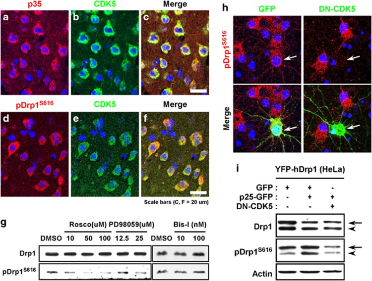
Figure 3.
Phosphorylation of Drp1S616 is mediated by CDK5 in neurons. Immunohistochemical labeling signals of p35 (a) and CDK5 (b), or pDrp1S616 (d) and CDK5 (e) in adult rat cerebral cortex. A merged image is shown in c and f. Nuclei were counterstained with Hoechst 33342 (blue). (g) Immunoblot of Drp1 and pDrp1S616 in cultured cortical neurons (DIV10) following the treatment with different concentrations of roscovitine, PD98059 and Bisindolylmaleimide I (Bis-I) for two hours. (h) Immunocytochemical labeling of pDrp1S616 (red) in cultured cortical neurons transfected with GFP (left panels) or DN-CDK5-GFP (right panels). Nuclei were counterstained with Hoechst 33342 (blue). Arrows indicate cells expressing GFP or DN-CDK5-GFP. (i) Immunoblot of Drp1 and pDrp1S616 in HeLa cells transfected with or without GFP, p25-GFP and DN-CDK5. Arrows indicate the bands derived from transfected YFP-hDrp1 proteins, while arrowheads indicate the bands derived from the endogenous Drp1.

