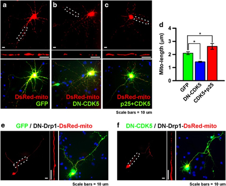
Figure 4.
Perturbation of CDK5 activity induces change on mitochondrial morphology. (a–c) Mitochondrial morphology of cultured cortical neurons in GFP- (a), DN-CDK5-GFP- (b), or CDK5-GFP and p25-transfected (c) groups at DIV10. Large, straightened mitochondrial morphology in white dotted boxes of a–c are shown at the bottom of each panel. (d) Average length of the mitochondria in each group. *P<0.05, n=10. (e, f) Mitochondrial morphology of cortical neurons cotransfected with DN-Drp1 and GFP (e) or DN-CDK5-GFP (f). Magnified images of the mitochondrial morphology in white dotted boxes are shown on the right side of each panel.

