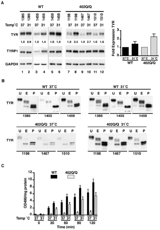
Figure 2. Temperature sensitive recovery of TYR activity in melanocyte strains.

(A) Endogenous protein expression of TYR and TYRP1 in genotyped primary melanocytes, WT (n=3) and 402Q/Q (n=3) incubated at 37°C and 31°C for a 24hr time period. Numbers represent normalised ratio of protein expression for TYR (37°C sample for each melanocyte strain set to 1) (lane 1,3,6,7,9,11 set to 1). A graph of this ratio represents a fold difference of TYR temperature sensitive protein expression between 37°C and 31°C in WT (n=3) and 402Q/Q (n=3) genotypes. Values indicate mean + SEM (n=3) of 3 different melanocyte strains of each genotype. GAPDH was used to determine protein loading. (B) Protein cell lysates of genotyped primary melanocytes, WT (n=3) and 402Q/Q (n=3) were untreated (U) or digested with the glycosidase EndoH (E) or PNGaseF (P). Samples were immunoblotted and probed with anti-TYR antibody to determine the extent of digestion. (C) The rate of L-DOPA oxidation in extracted cell lysates of primary melanocytes, WT (n=3) and 402Q/Q (n=3) at 37°C and 31°C was determined over a 2hr time course at 30min intervals. Values indicate mean + SEM (n=3) of 3 different melanocyte strains of each genotype normalised to total soluble protein.
