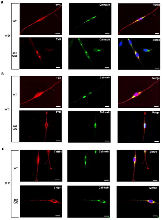
Figure 3. Localization of TYR in 402Q/Q melanocytic cells is temperature dependent.

The images show immunofluorescent staining of WT (QF 1459) and homozygous variant 402Q/Q (QF 1510) melanocyte strains grown on coverslips at 37°C (A and C) or shifted to 31°C for 24h (B). In panels A and B, cells grown at indicated temperature were fixed and stained with mouse TYR-Alexa-594 (red) and rabbit calnexin-Alexa-488 (green). In panel C, cells grown at 37°C were fixed and stained with mouse TYRP1-Alexa-594 (red) and rabbit calnexin-Alexa-488 (green). Left to right with the antibody indicated in the upper right: TYR or TYRP1 (red), calnexin (green) and an overlay of the coloured images also including DAPI (blue). Yellow indicates colocalization between TYR/TYRP1 and calnexin. Scale bars in white represent 50μm.
