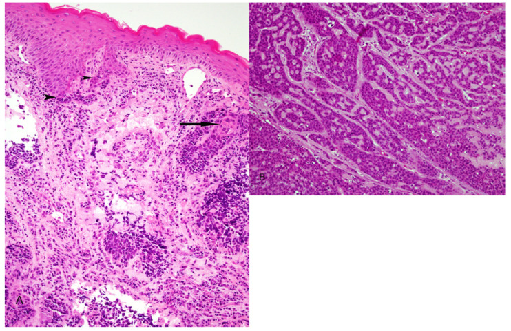
Figure 3.
Basaloid squamous cell carcinoma. A) Illustrates biphasic pattern with conventional dysplastic squamous surface component associated with basaloid elements (arrow heads) and conventional squamous cell carcinoma intimately associated with basaloid component (arrow). B) Closely packed basaloid cells forming a "jigsaw puzzle" appearance. Microcystic cribriform-like pattern is also observed.

