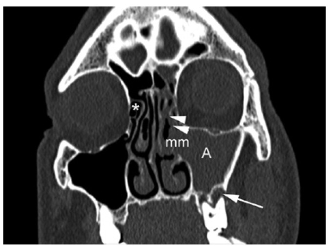
Figure 2.
CT scan (coronal plane) corresponding to a 49 years old man patient showing a left odontogenic maxillary sinusitis extended to anterior ethmoid sinus. There are a diffuse opacification of the maxillary sinus (A) and anterior ethmoid sinus (arrowheads). The middle meatus (mm), where are situated the ostium of maxillary and anterior ethmoid sinus, is compromised by the inflammatory process. The anterior ethmoid sinuses on the right side are normal (*). The arrow indicates the location of a periapical lesion (abscess) adjacent to the sinus floor.

