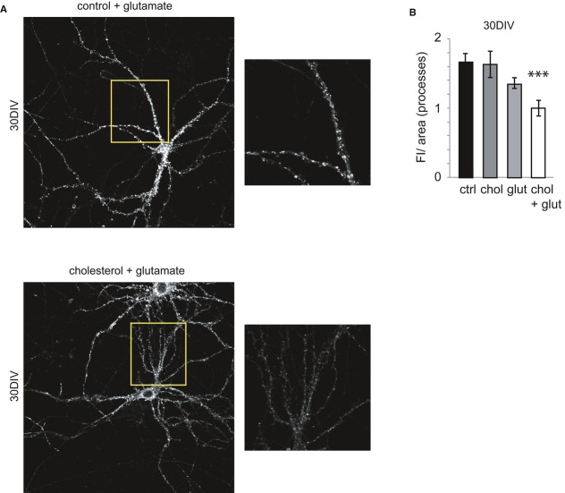
Figure 4. Glutamate addition decreases the level of surface AMPARs in cholesterol-replenished 30 DIV neurons.
- The surface AMPAR staining decreased in cholesterol-replenished 30 DIV neurons after a 10-min incubation with glutamate. The boxes on the right correspond to high-magnification images of the regions indicated in the insets.
- The bar plot shows the quantification of the fluorescence intensity (FI)/area (mean ± s.e.m., relative to the cholesterol + glutamate condition) in the processes of control and cholesterol-replenished neurons. No differences in the FI/area values were found in control, glutamate-stimulated (control = 1.67 ± 0.12, glutamate = 1.35 ± 0.07; P = 0.053; n = 3) or cholesterol-replenished unstimulated neurons (control = 1.67 ± 0.12; cholesterol = 1.63 ± 0.19; P = 0.87; n = 3). Stimulation of cholesterol-replenished neurons (chol + glut) provoked a significant decrease in the surface GluA2 staining (control = 1.67 ± 0.12; cholesterol + glutamate = 1.00 ± 0.12; P = 0.0018; n = 3). The data were compared using Mann-Whitney non-parametric t-test.

