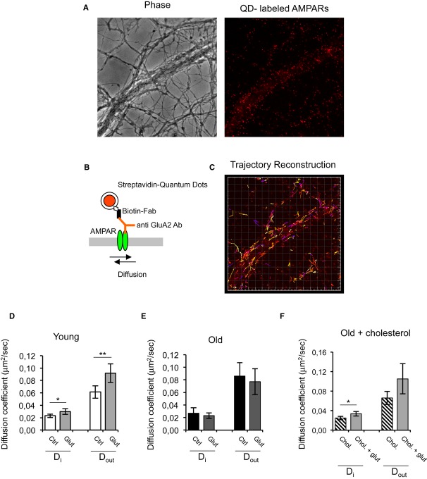
Figure 5. Lateral diffusion of GluA2-AMPARs is altered in low-cholesterol hippocampal neurons after LTD induction.
- A Phase contrast and fluorescence images showing the processes of hippocampal neurons where GluA2 subunits were labeled with quantum dots.
- B GluA2-AMPARs were labeled using an anti GluA2 antibody and a biotinylated anti-mouse Fab fragment conjugated to streptavidin-coated quantum dots.
- C Image showing the reconstructed trajectories of individual GluA2-AMPARs.
- D, E Glutamate stimulation increases the diffusion of synaptic and extra-synaptic AMPARs in 15 DIV but not 30 DIV neurons. 15DIV: Di control = 0.023 ± 0.0028 μm2/s, Di glut = 0.0298 ± 0.0045 μm2/s (P = 0.0201), Dout control = 0.0613 ± 0.0099 μm2/s, Dout glut = 0.0917 ± 0.0149 μm2/s (P = 0.0054). 30 DIV: Din control = 0.0269 ± 0.0085 μm2/s, Din glut = 0.0231 ± 0.0044 μm2/s (P = 0.4048) and Dout control = 0.086 ± 0.0213 μm2/s, Dout glut = 0.0771 ± 0.0207 μm2/s (P = 0.5228).
- F The addition of cholesterol to 30 DIV neurons restores the response of synaptic AMPARs to glutamate. Din chol = 0.0250 ± 0.0035 μm2/s, Din chol + glut = 0.0337 ± 0.0047 μm2/s (P = 0.0254).

