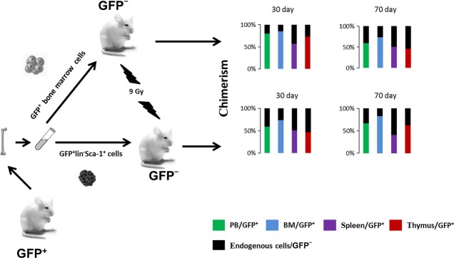
Fig. 1.

Model for the analysis of chimerism after cell transplantation. Our experimental model of C57BL/6-Tg(CAG-EGFP)C14-Y01-FM131Osb mice as donors of GFP+ bone marrow (BM) or GFP+lin− Sca-1+ cells and C57Bl6/J mice as recipients (GFP−). Recipient animals were exposed to 9 Gy whole-body irradiation. Suspensions of GFP+ bone marrow cells (5 × 10⁶/ml) or GFP+lin−Sca-1+ cells (3 × 10⁴/ml) were transplanted by i.v. injection into recipient animals 3 hrs after irradiation. Flow cytometry analysis (FCA) of all GFP+ cells (%) was performed on days 30 and 70 after cell transplantation. We detected chimerism GFP+ cells (%)/GFP− cells (%) in the peripheral blood (PB), bone marrow (BM), spleen and thymus.
