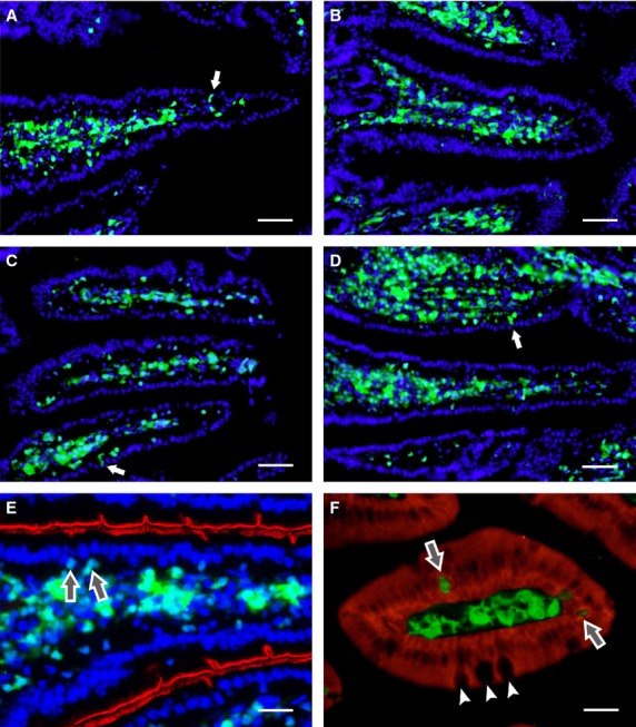
Fig. 4.

Histological analysis of cell engraftment in the small intestine. By day 30 after transplantation, the stroma of intestinal villi are richly colonized with green fluorescent protein (GFP+) cells derived from grafted bone marrow cells (A) or lin− cells (B). GFP+ cells remain in the core of the villi for at least 70 days after the transplantation of unseparated bone marrow cells (C and E) or lin− cells (D). A few GFP+ cells are closely associated with, or incorporated into, the epithelial lining (arrows). The brush border of enterocytes is indicated by red pseudocolour derived from the histochemical activity of intestinal alkaline phosphatase (E). Enterocytes express pancytokeratin (red), while GFP+ cells, including those incorporated in the epithelial lining (arrows), do not. Goblet cells (small arrows) are GFP− (F). Cell nuclei were counterstained with DAPI; scale bar: A–D 100 μm, E 25 μm, F 20 μm.
